Twitter
Home
About Us
About ASSCR
ASSCR rising star award
Committee Members
Annual Reports
ASSCR Committee Election
About Stem Cells
What are Stem Cells?
Research Centres and Networks in Australia
Organisations Around the World
For Patients
Meetings & Events
Public Forum: Stem Cell Research – Now and in the Future
Upcoming Events
Australia
International
Past Events
2023 Conference Photo Gallery
2022 Conference Photo Gallery
ASSCR Meetings
Stem Cell Stories Exhibition
Membership
News
Science Meets Parliament 2023
Science Meets Parliament 2023 blog
Science Meets Parliament 2024
Contact Us
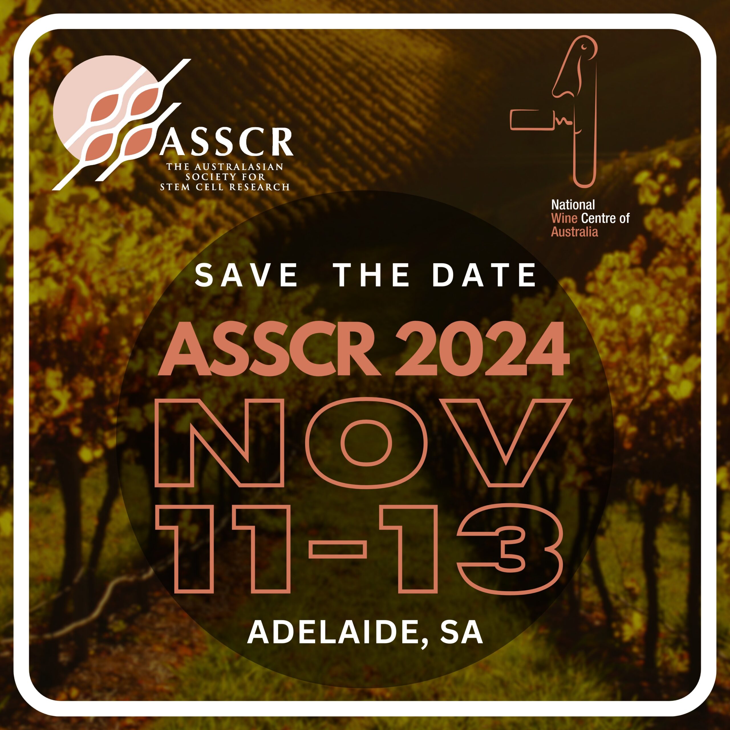
Save the date
Follow @the_ASSCR
Tweets by the_ASSCR
Recent Past ASSCR Events in Australia
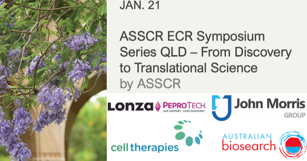
ASSCR ECR Symposium Series: From Discovery to Translational Science
Date:
January 21, 2021
Time:
12:00 am - 12:00 am
Location:
Abel Smith Lecture Theatre University of Queensland (Building 23) St Lucia, QLD 4072
More info
ASSCR Annual Meetings | Australia
Search for more Stem Cell Events
Copyright © 2018 The Australasian Society for Stem Cell Research (ASSCR). All rights reserved.